
Supine Arthroscopy for Humeral Osteochondral Lesion
Lee Hunter MD, MBA (Hunter Medical Founder and Chief Medical Officer)
March 2020
The advent of arm positioning systems designed specifically for elbow surgery has greatly simplified supine elbow arthroscopy.
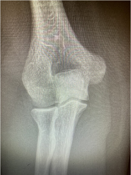
39 YOF With Large Humeral Osteochondral Lesion
The patient presented complaining of chronic lateral right elbow pain for 4 months. She had received one intra-articular cortisone injection elsewhere which provided no relief. She denied any traumatic event. Her medical history was notable only for diet-controlled diabetes and a seizure disorder, controlled with medication. Physical exam was notable for lateral elbow joint line tenderness, an effusion, 25-degree elbow flexion contracture, and flexion to 130 degrees. Forearm rotation was mildly uncomfortable but supple. Her orthopaedic exam was otherwise unremarkable. Preoperative plain films and MRI are shown below.


The Procedure
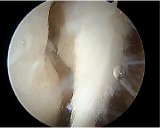
The patient was positioned supine utilizing the ElbowLOC® Arm Positioning System. Standard arthroscopic portals were created. The fragment was floating loose, contained nonviable subchondral bone and was removed. Loose necrotic bone was debrided from the base of the lesion which was then treated with standard microfracture technique. The large size of the lesion, it’s relatively centered location on the capitellum, and the lack of lateral wall support made it a poor choice for an osteochondral graft.


The ElbowLOC® Supine Positioner is an ideal tool for elbow arthroscopy. It enables incremental, easy adjusting of intraoperative elbow position while allowing circumferential working space for arthroscopic tools.
This case is another great example of how the ElbowLOC® simplifies and accelerates procedural times for many elbow surgeries and makes surgery easier for the surgeon, patient, and staff!