
Lateral Ulna Collateral Ligament Reconstruction
Lee Hunter MD, MBA (Hunter Medical Founder and Chief Medical Officer)
July 2017
Chronic lateral elbow instability can be treated successfully with a lateral ulna collateral ligament reconstruction. Supine patient positioning is ideal for these procedures.
45 YO RHDM With Chronic Lateral Elbow Instability
The patient had chronic lateral elbow instability, with a history of prior open tennis elbow release and more than 10 years of activity-related lateral elbow pain. Conservative treatment included activity modification and repeated supervised physical therapy courses over several years. Routine x-rays were unremarkable. Clinical exam revealed a palpable defect in the posterolateral elbow soft tissue, along with exam findings consistent with lateral instability. MRI revealed at least a partially intact wrist extensor origin, but no identifiable LUCL tissue.
The Procedure
The patient was positioned supine utilizing the ElbowLOC®Arm Positioning System. A posterolateral approach was made. An autograft palmaris longustendon graft double looped was utilized. The graft was secured proximally and distally with PEEK interference screws, and the sutures additionally woven through surrounding capsular tissue. Postoperatively, a controlled motion brace with a 20-degree extension block was utilized for 3 weeks, then unlocked. At a year follow-up, patient had a 5-degree flexion contracture, otherwise full motion with subjectively normal strength and no pain.
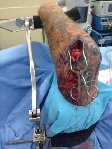


The ElbowLOC®Supine Positioner markedly simplified and expedited this case by stabilizing the humerus vertically while allowing excellent visualization of and access to the joint. With the elbows table, proper seating/congruency of the proximal ulna within the trochlear groove is assured. This case is another great example of how the ElbowLOC®simplifies and accelerates procedural times for many elbow surgeries and makes surgery easier for the surgeon, patient, and staff!