Supine Elbow Arthroscopy for Osteochondritis Dissecans In a Throwing Athlete
Lee Hunter MD, MBA (Hunter Medical Founder and Chief Medical Officer)
September 2016
14-year-old RHD Baseball Player with Osteochondritis Dissecans
Osteochondritis Dissecans of the elbow presents in various stages of severity, which govern the treatment. Treatment options for unstable osteochondral fragments include surgical stabilization if possible or removal of loose bodies and debridement with microfracture of the base versus osteochondral grafting. The size and location of the lesion on the capitellum also influence the treatment method. Sometimes the location of the lesion and the presence of the overlaying radial head can make treatment difficult. The case illustrated below is a great example.
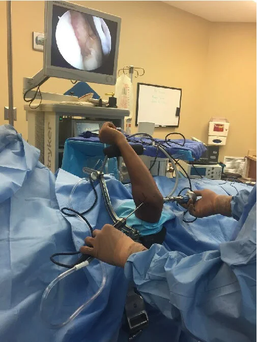
The Case
The patient presented with the primary complaint of activity related lateral elbow pain for more than a year. When working out recently, he felt a painful click, and since then has had stiffness and more discomfort. Radiographs revealed a large, displaced osteochondral lesion with the bed measuring roughly 14 x 14 mm. Arthroscopy was performed using the ElbowLOC® in supine positioning mode. This allows very good arthroscopic or open visualization of all parts of the distal humeral articular surface. Additionally, the surgeon maintains complete control of the forearm and can “lock” the elbow statically in the exact position needed, without relying on multiple surgical assistants and the inevitable drifting and motion that brings. The lesion was treated with removal of the loose bodies and debridement/microfracture of the base. Its relatively anterior position on the capitellum, made it more a bit more challenging to visualize and access than the relatively posterior centered lesions. The attached photos show how the ElbowLOC® helped simplify and expedite this case. Elbow hyperflexion is easily accomplished by placing the patient’s hand on his chest, which allows visualization and treatment of the posterior aspect of the capitellum. Static elbow extension was facilitated by using the ElbowLOC® Reducer/Distractor component instead of the standard wrist Support. This maximizes the surgeon’s ability for static elbow joint extension and enables visualization and treatment of the anterior aspect of the capitellum. This may be particularly important during elbow arthroscopy on patients of shorter stature, as it allows the ElbowLOC® Supine Arm to remain fully seated in the Base Support and creates maximum working area for arthroscopic instruments.